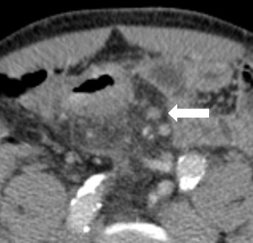
Fig. 3.

Transaxial reformation (5 mm section post iv contrast portal phase) CT image illustrating the apperance of a 7 × 7 mm mesocolic lymph node (white arrow) in region 2 with irregular outer border and internal heterogeneity in a patient with a pT3 tumour in the sigmoid colon with 1 metastatic lymph nodes out of 16 harvested at histopathology
