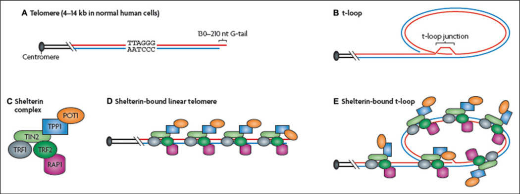
Figure 1.
Structure of telomeres. A, Vertebrate telomeres contain repetitive DNA with the sequence (5′-TTAGGG-3′). Most of this DNA is double stranded, apart from the terminus, which consists only of the TTAGGG (or “G-rich”) strand. B, The telomere can fold back on itself, and the single-stranded terminus can invade duplex telomeric DNA. This results in the formation of a telomeric loop (t-loop). C, Telomeric DNA is bound by the 6-unit shelterin complex. D and E, Two shelterin proteins (telomeric repeat-binding factor 1 [TRF1], also known as TERF1 and TRF2, also known as TERF2) bind directly to double-stranded telomeric DNA, and one (protection of telomeres 1 [POT1]) binds to single-stranded telomeric DNA directly. POT1, TPP1 (also known as ACD), TRF1-interacting nuclear protein 2 (TIN2, also known as TINF2), and repressor activator protein 1 (RAP1; also known as TERF2IP) also interact indirectly with double-stranded telomeric DNA through their interactions with other shelterin proteins. Reprinted by permission from Macmillan Publishers Ltd.. Cesare, A. J., & Reddel, R. R. (2010). Alternative lengthening of telomeres: Models, mechanisms, and implications. Nature Reviews Genetics, 11, Fig. 1, p. 320.

