
Figure 3.
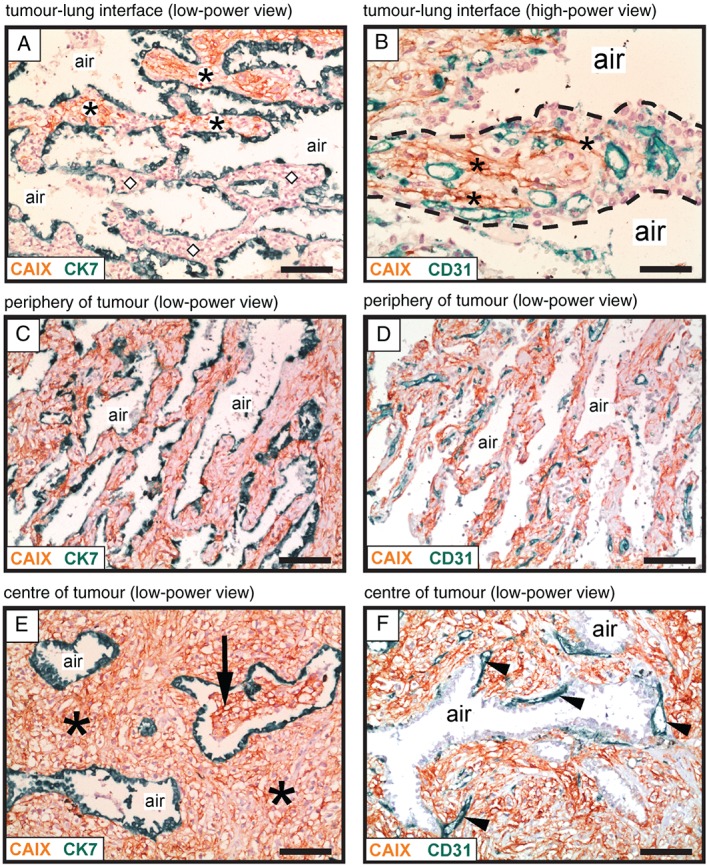
Vessel co‐option occurs in the interstitial growth pattern of human lung metastases. Immunohistochemical analysis of a renal cancer lung metastasis with an interstitial HGP, illustrating growth of cancer cells within the alveolar walls. Staining for CAIX (brown) was used to detect cancer cells, in combination with either CK7 staining (green) to detect pneumocytes (A, C, E) or CD31 staining (green) to detect blood vessels (B, D, F). (A) Tumour–lung interface: alveolar walls filled with cancer cells are present at the top of the image (asterisks), whereas tumour‐free alveolar walls of the normal lung are present below (diamond symbols). (B) High‐power view of an alveolar wall (delineated with a dashed line). Asterisks indicate cancer cells that are infiltrating around pre‐existing alveolar capillaries. (C, D) The area just behind the tumour–lung interface: the alveolar walls are now completely filled with cancer cells. The intervening alveolar air spaces remain intact. (E, F) The centre of the metastasis. In (E), asterisks indicate cancer cells that are filling the expanded alveolar walls, while the intervening alveolar air spaces remain intact. The arrow indicates an alveolar air space that has become partially filled with cancer cells. In (F), arrowheads indicate blood vessels that are closely associated with the abluminal side of an alveolar air space. Alveolar air space: air. Scale bars: 100 µm (A, C, D, E, F) and 50 µm (B).