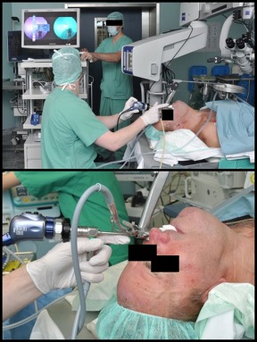
Figure 1.

Intraoperative setting. Standard panendoscopy was performed, including suspension laryngoscopy. Rigid near‐infrared (NIR) with indocyanine green (ICG) endoscopy was performed directly after injection of the ICG. A split screen allowed observation of white light image and fluorescence image at the same time. [Color figure can be viewed at wileyonlinelibrary.com.]
