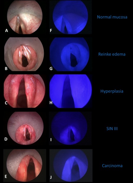
Figure 2.

Endoscopic examples of near‐infrared (NIR) with indocyanine green (ICG) finding in different types of mucosal head and neck lesions in the larynx. White light image on the left side (A–E) and corresponding NIR ICG image on the right side (F–J). Normal mucosa (AB) and Reinke edema (BG) only showed ICG positivity in the submucosal vessels. The mucosal hyperplasia on the right anterior vocal cord (CH) was completely ICG‐negative, even sparing ICG‐positive vessels. The severe dysplasia of the right vocal cord (squamous intraepithelial neoplasia [SIN] III, DI) and the squamous cell cancer of the left anterior vocal cord (T1 glottic cancer, EJ) showed a diffuse ICG positivity and retention of ICG. [Color figure can be viewed at wileyonlinelibrary.com.]
