Figure 2.
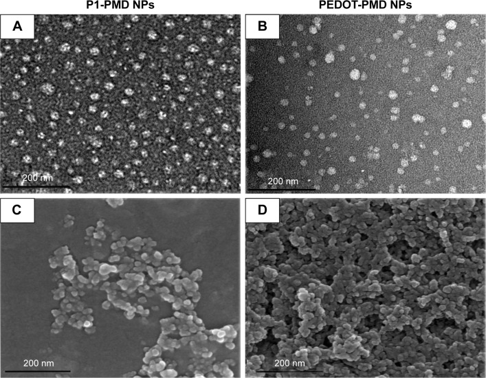
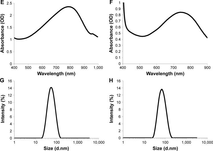
Characterization of P1-PMD and PEDOT-PMD NPs.
Notes: Transmission electron microscopy (A, B) and scanning electron microscopy (C, D) microscopy images of NPs. Absorption spectra (E, F) and dynamic light scattering size distribution of (G, H) NPs.
Abbreviations: NPs, nanoparticles; P1, poly(diethyl-4,4′-{[2,5-bis(2,3-dihydrothieno[3,4-b][1,4]dioxin-5-yl)-1,4-phenylene]bis(oxy)} dibutanoate); PEDOT, poly(3,4-ethy-lenedioxythiophene); DBSA, 4-dodecylbenzenesulfonic acid; PSS-co-MA, poly(4-styrenesulfonic acid-co-maleic acid); P1-PMD, P1:PSS-co-MA:DBSA; PEDOT-PMD, PEDOT: PSS-co-MA:DBSA.


