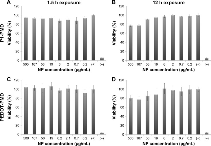
Figure 5.
Cytocompatibility of P1-PMD and PEDOT-PMD NPs in MDA-MB-231 breast cancer cells determined with the MTT assay.
Notes: Data shown as percent viability relative to positive control (+), which consists of cells cultured in NP-free media. Negative control (−) represents cells killed with methanol. Error bars represent the standard deviation between replicates (n=6). Cells exposed to (A) P1-PMD NPs for 1.5 h, (B) P1-PMD NPs for 12 h, (C) PEDOT-PMD NPs for 1.5 h, and (D) PEDOT-PMD NPs for 12 h.
Abbreviations: MTT, 3-(4,5-dimethylthiazol-2-yl)-2,5-diphenyltetrazolium bromide; NP, nanoparticle; P1, poly(diethyl-4,4′-{[2,5-bis(2,3-dihydrothieno[3,4-b][1,4]dioxin-5-yl)-1,4-phenylene]bis(oxy)} dibutanoate); PEDOT, poly(3,4-ethylenedioxythiophene); DBSA, 4-dodecylbenzenesulfonic acid; PSS-co-MA, poly(4-styrenesulfonic acid-co-maleic acid); P1-PMD, P1:PSS-co-MA:DBSA; PEDOT-PMD, PEDOT:PSS-co-MA:DBSA.

