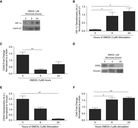
Figure 3.
Stabilization of HIF-1α in normoxia by dimethyloxaloylglycine (DMOG) results in CD55 down-regulation in SAECs. (A) DMOG-treated (1 μM) SAECs were probed for HIF-1α protein expression. Lamin β1 was used as internal loading control. (B) Densitometry analysis shows HIF-1α induction within 24-hour DMOG (1 μM)-treated SAECs compared with cells with no DMOG. Values represent means (±SEM); n = 3; *P < 0.05 versus nontreated cells (0 hour controls); one-way ANOVA with Bonferroni posttest. (C) CD55 transcripts were down-regulated in 6-hour DMOG (1 μM)-treated SAECs compared with the nontreated cells (0 hour controls). Data represent means (±SEM); n > 3; **P < 0.01 versus 0 hour (nontreated cells); one-way ANOVA with Bonferroni posttest. (D) CD55 was probed for in DMOG (1 μM)-treated SAECs. Lamin β1 was used as internal loading control. (E) Densitometry analysis shows CD55 down-regulation by 24 hours of DMOG (1 μM)-treated SAECs compared with the nontreated cells (0 hour controls). Values represent means (±SEM); n = 3; **P < 0.01 versus nontreated cells (0 hour controls); one-way ANOVA with Bonferroni posttest. (F) CA9 transcripts were up-regulated in DMOG (1 μM)-treated SAECs compared with nontreated cells. Data represent means (±SEM); n = 3; *P < 0.05, or **P < 0.01 versus nontreated cells (0 hour controls); one-way ANOVA with Bonferroni posttest.

