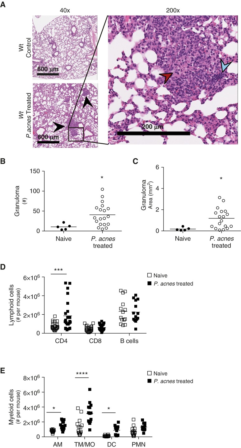
Figure 1.
Generation of early pulmonary granuloma model. Wild-type (Wt) mice were administered 109 viable Propionibacterium acnes intratracheally and were killed after 9 days to generate a mouse model of sarcoid-like granuloma formation. Naive Wt mice were used as control mice. (A) Naive (top left) and P. acnes–administered Wt mouse lungs (bottom left) were removed, fixed, embedded, and examined for granulomas (black arrowheads) via hematoxylin and eosin staining at low magnification. Scale bars: 600 μm. At higher magnification in P. acnes–administered mice (right), epithelioid macrophages (red arrowhead) and lymphoid cells (blue arrowhead) can be observed. Scale bar: 200 μm. Granuloma (B) numbers and (C) area were quantified using pattern recognition software Spatially Invariant Vector Quantization (SIVQ) from three tissue sections 200 μm apart; each individual data point represents the average of those measurements per mouse. (D and E) Naive and P. acnes–administered lungs were also perfused with phosphate-buffered saline, digested into single-cell suspensions, analyzed for cell type composition via flow cytometry, and gated as described in Materials and Methods. The number of total viable (D) lymphoid cells and (E) myeloid cells is shown. Lymphoid cells are CD4+, CD8+ T cells, and B cells; myeloid cells are AM, TM/MO, DC, and PMN. Statistical significance was determined using unpaired t tests (B and C) and multiple t tests (D and E). *P < 0.05, ***P < 0.001, and ****P < 0.0001. AM, alveolar macrophages; DC, dendritic cells; PMN, neutrophils; TM/MO, tissue macrophages/monocytes.

