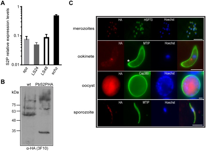
Fig 2. Expression and localisation of PbS2P.
(A) Relative expression levels of PbS2P as determined by qRT-PCR from cDNAs of schizonts (schz), sporozoites (spz), 24h liver stages (LS24) and 48h liver stages (LS48). Transcript levels were normalised to PbHSP70 and GFP. (B) Western blot analysis of PbS2P-HA whole protein extract from purified schizonts of transgenic PbS2P-HA parasites using an α-HA antibody. PbS2P-HA migrates at 35kDa. (C) Immunofluorescence analysis (IFA) of PbS2P-HA merozoites, ookinete, oocyst, and salivary gland sporozoites using α-HA (3F10) for detection of PbS2P (red) and Hoechst stain for the nucleus (blue). For delineation of parasites the following antibodies (green) were used: α-HSP70, schizonts/merozoites; α-MTIP, ookinete and sporozoite; α-PbCap380, oocyst. Prominent localisation of PbS2P in proximity to the nucleus is present in all invasive stages. Star, apical end of ookinete. Scale bar 5 μM.

