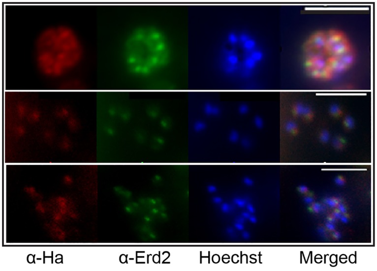
Fig 3. PbS2P shows partial co-localisation with the cis-Golgi marker ERD2.

(A) Double labelling IFA of P. berghei schizont cultures using α-HA (3F10) for detection of PbS2P (red) and α-ERD2 as a Golgi marker (green) showing partial, or in some cases complete, co-localisation. Nuclei are stained with Hoechst (blue). Scale bar 5 μM.
