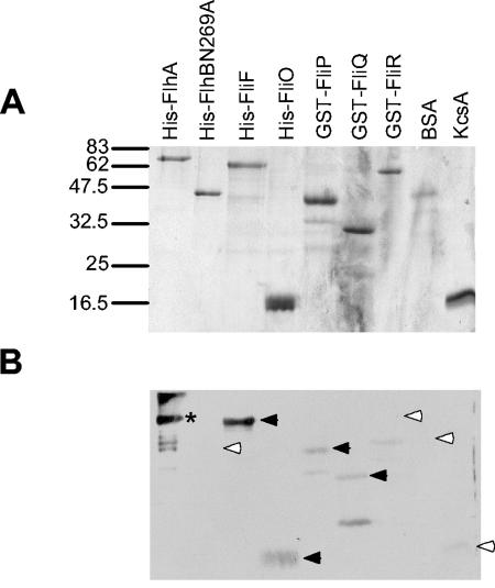
FIG. 3.
Affinity blotting of membrane components of the export apparatus versus purified His-FlhA. (A) Coomassie blue-stained gel of purified membrane export apparatus components. Positions of molecular weight standards are shown on the left in thousands. (B) Affinity blot of the same samples with 10 μg of His-FlhA and anti-FlhAc antibody per ml. Significant signals are indicated by black arrowheads; absence of signal and weak or insignificant signals are indicated by white arrowheads. The asterisk denotes an anti-FlhA-reactive band that represents direct antibody binding to FlhA.

