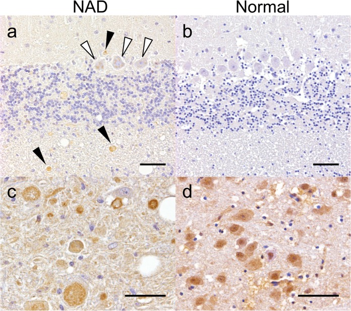
Fig 4. iPLA2β immunohistochemistry of Case 1 (a, c) and a normal (b, d) canine brain.
(a) Spheroids in the cerebellum showed faint granular expression (black arrowheads). Cytoplasmic granular expression was also observed in the Purkinje cells (white arrowheads). (b) The expression was very weak in the cerebellum of the control dog. In the brainstem (c, d), intense cytoplasmic and nuclear expression was detected in the neurons of both the NAD and control dogs. Axonal spheroids in the neuropil of the brainstem of Case 1 showed an intense granular expression (arrowheads). Bar = 50 μm.

