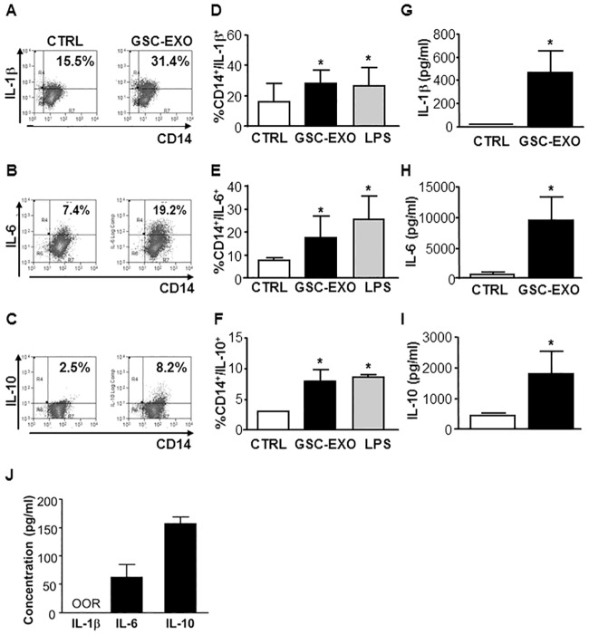
Fig 5. GSC-derived exosomes stimulate IL-1β, IL-6 and IL-10 production in unstimulated CD14+ monocytes within PBMC population.
Unstimulated PBMCs were incubated in the absence (white column, CTRL) or presence (black column, GSC-EXO) of GSC-derived exosomes. Incubation with LPS (square column) was used as monocyte stimulation positive control. Cells were surface stained with anti-CD14 and then stained to detect an intracellular level of IL-1β, IL-6 and IL-10 by flow cytometry. (A-C) Representative FACS plot of the intracellular staining is shown by the indicated percentage of CD14+/IL-1β+, CD14+/IL-6+ and CD14+/IL-10+ positive cells, respectively. (D-F) The mean of the experiments is shown (n = 6); bars, SD; *, significantly different from the control; P<0.05. Supernatants of unsorted unstimulated PBMCs incubated in the absence (white column) or presence of GSC-derived exosomes were harvested after 48 hours and used for a cytokine assay with the Bio-plex cytokine assay system. (G-I). As a control, cytokine concentration was also tested on a medium added with isolated exosomes (J). Concentration of IL-1β, IL-6 and IL-10, respectively, are expressed as pg/ml. Columns, mean (n = 6); bars; SD; *, significantly different from the control; P<0.05.

