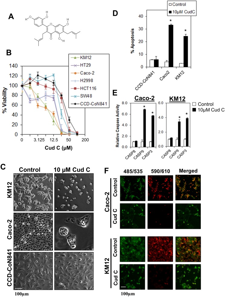
Fig 1. Cudraflavone C induces tumor-specific cell death in colorectal cancer cells.
(A) Chemical structure of Cud C. (B) KM12, HT29, Caco-2, HCC2998, HCT116 and SW48 colorectal cancer cells were exposed to various concentrations of Cud C for 72 hours. Cell viability was recorded using CellTitre Glo® luminescence assay. (C) KM12, Caco-2 and CCD 841 CoN were treated with 0.1% DMSO (control) or 10μM Cud C (Cud C) for 72 hours followed by microscopy analysis (×100 magnification). (D) Apoptotic cell death in KM12, Caco-2 and CCD 841 CoN cells was quantified using Annexin V/7-AAD flow cytometry at 72 hours following treatment. (E) Caspase activities in KM12 and Caco-2 cells were assessed by Caspase Glo® assay at 72 hours following treatment. (F) 10μM Cud C induced mitochondrial membrane depolarization. Caco-2 and KM12 cells stained with JC-1 at 72 hours after treatment with Cud C. The green dye represents JC-1 monomers in cytoplasm while the red dye represents JC-1 aggregates in nucleus. Cells were observed under fluorescence microscope (×100 magnification). All data represent the mean ± s.d. from at least three independent experiments. Symbol “*” presents the statistical significance concluded from Student’s independent t-test with p-value ≤0.05.

