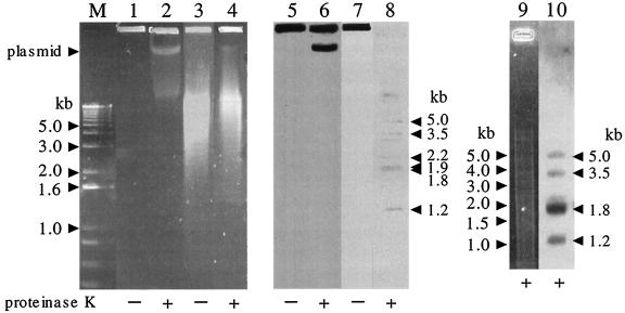
FIG. 1.
Analysis of RHA1 telomere fragments. RHA1 cells were lysed in an agarose plug with (+) or without (−) proteinase K treatment. Agarose plugs containing RHA1 DNA were subjected to PFGE directly (lanes 1, 2, 5, and 6) or after PstI digestion (lanes 3, 4, 7, and 8). Electrophoresis was conducted for 6 h with a voltage of 6 V/cm and a pulse time that was increased from 2 to 10 s as the electrophoresis progressed. Lanes M to 4 and 9 were stained with ethidium bromide. Lanes 5 to 10 represent Southern blots with a probe derived from the right telomere of pRHL3. The experiment shown in lanes 9 and 10 was performed by using conditions of higher stringency. Lane M, 1-kb plus DNA ladder size marker (Invitrogen, Carlsbad, Calif.). The position of intact linear plasmid DNA containing pRHL3 is indicated on the left. The estimated sizes of the fragments detected by hybridization are indicated on the right.

