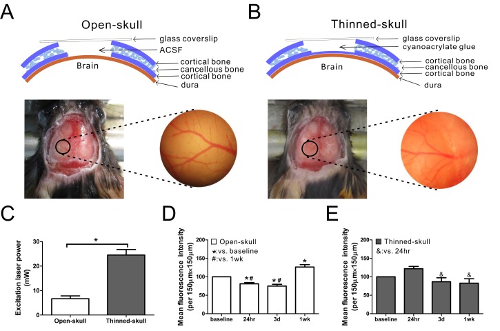
Fig 1. Schematic diagram and image analysis of open-skull technique and thinned-skull techniques for in vivo imaging of astrocytes in GFAP-GFP mice.
(A,B) The mouse skull consists of two thin layers of cortical bone and a thick layer of cancellous bone. In the open-skull technique, all three layers of skull are removed and the exposed dura is coated with ACSF and covered with a glass coverslip over the skull window (A). In the thinned-skull technique, the skull is carefully thinned to the inner cortical bone to about 20 μm in thickness. The thinned skull is coated with a layer of cyanoacrylate glue and covered with a glass coverslip over the skull window (B). In both techniques, a round area of skull (~2 mm in diameter, marked with black circle) over the left somatosensory neocortex is removed (A) or thinned (B). Vasculature images captured after surgery help to ensure repeated observation from the same areas. (C) About 7 mW excitation laser power was needed for good image quality in the open-skull technique but much higher laser power (~25 mW) was required in thinned-skull technique to provide a similar image intensity (*p<0.05 by t-test, n = 6 per group). (D) In the open-skull technique, mean fluorescence intensity decreased 24 hr and 3 d after surgery but increased at 1wk (*,# p<0.05 by one-way ANOVA with Tukey post test, n = 6 per group). (E) In contrast, no significant change of mean fluorescence intensity was observed in the thinned-skull technique over time compared with baseline (p>0.05), although there was a slight decrease in intensity at 3 d and 1 wk compared to 24 hr (& p<0.05 by one-way ANOVA with Tukey post-test, n = 6 per group).

