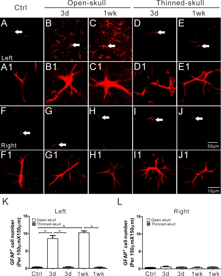
Fig 4. Immunohistochemical staining of GFAP in fixed tissue sections following open-skull and thinned-skull surgery in GFAP-GFP transgenic mice.
Expression of GFAP (red) is a prototypical immunohistochemical marker of reactive astrocytes. GFAP positive astrocytes increased massively on the surgery side (left side) of the neocortex at 3 d and 1 wk after open-skull surgery (B, C) but not after thinned-skull surgery (D, E). Minimal GFAP positive astrocytes occurred on the contralateral side (right side) (G-J) and in control mice (A, F). The arrows in figures A-J denote the astrocytes that are enlarged as in figure A1-J1, respectively. Summarized data (K, L) indicate significantly more GFAP positive astrocytes in the left neocortex at 3 d and 1 wk in open-skull mice than in thinned-skull mice or in control mice (K), with no significant difference in the right neocortex (L). *p<0.05, by one way ANOVA with Tukey's post-test (n = 6 per group).

