
Figure 3.
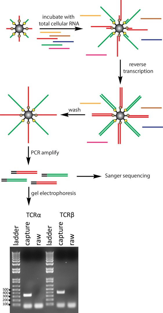
RNA from lysed T cells was incubated with capture beads having sequences complementary to the constant region of TCRα and TCRβ. Captured RNA was reverse transcribed and PCR amplified, and then it was resolved using gel electrophoresis and visualized by ethidium bromide staining. PCR reactions from the capture beads show amplification of TCRα and TCRβ sequences. As a control, all of the above steps were performed using raw beads (no oligos attached) and show no PCR product. The size (in base pairs) of the smallest five bands in the DNA ladder are indicated to the left of the first gel image.