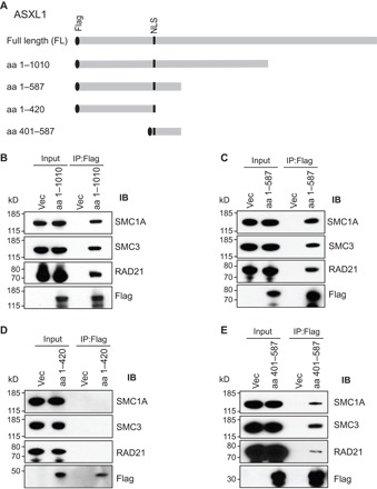
Fig. 2. Mapping the region of ASXL1 that mediates its binding to the cohesin complex.

(A) Schematic diagram of the full-length (FL) ASXL1 and the truncated variants of Asxl1 [amino acids (aa) 1 to 1010, 1 to 420, 1 to 587, and 401 to 587]. Binding affinity was determined by the pull-down efficiency of IP with anti-FLAG and Western blotting with cohesin antibodies. NLS, nuclear localization signal. (B to E) Western blotting analysis of nuclear fractions and anti-FLAG immunoprecipitates from pcDNA3.1+, or each truncated ASXL1 transfected HEK293T cells using antibodies against FLAG, SMC1A, SMC3, or RAD21.
