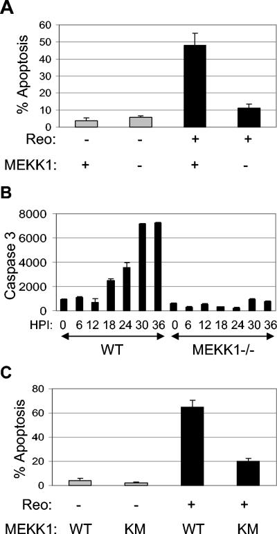
FIG. 1.
Reovirus-induced apoptosis is inhibited in MEKK1−/− cells and in cells expressing MEKK1KM. MEKK1−/− cells were infected with reovirus (MOI of 10; black bars) or were mock infected (gray bars), and the percentage of cells containing apoptotic nuclei at 48 h p.i. was determined (A). The graph shows the mean percentage of apoptotic cells from three independent experiments. Error bars represent standard errors of the mean. Reovirus-induced activation of caspase 3 was also determined for MEKK1−/− cells and controls (WT) at various times p.i. by fluorogenic substrate assay (B). The graph shows the mean fluorescence (arbitrary units) from three wells of an individual experiment, which represents caspase 3 activity, and is representative of three separate experiments. Cells expressing MEKK1KM and controls were infected with reovirus (MOI of 100; black bars) or were mock infected (gray bars), and the percentage of cells containing apoptotic nuclei at 48 h p.i. was determined (C). The graph shows the mean percentage of apoptotic cells from three independent experiments. Error bars represent standard errors of the mean.

