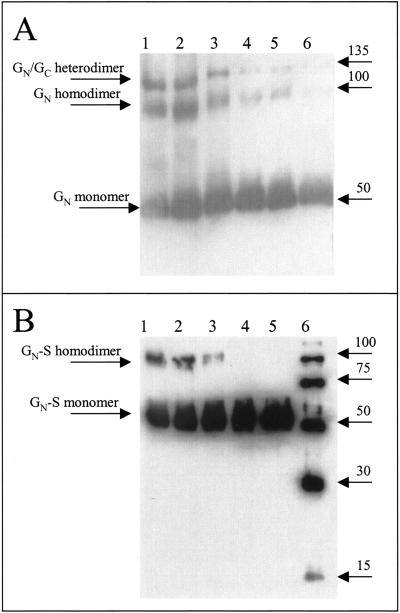
FIG. 6.
Analysis of GN and GN-S dimerization. Increasing amounts of β-ME were added to TSWV purified from infected plants (GN) or to GN-S. Proteins were detected by Western blotting. (A) Purified TSWV detected with GN MAb. Lane 1, no β-ME and sample was not boiled; lane 2, no β-ME; lane 3, 0.1% β-ME; lane 4, 1.0% β-ME; lane 5, 2.5% β-ME; and lane 6, 5% β-ME. (B) Purified GN-S protein was detected with a six-His MAb. Lane 1, no β-ME and sample was not boiled; lane 2, no β-ME; lane 3, 0.1% β-ME; lane 4, 1.0% β-ME; lane 5, 2.5% β-ME; and lane 6, six-His-tagged molecular weight marker.

