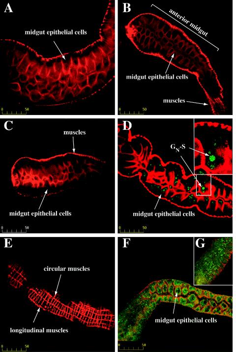
FIG. 7.
In vivo binding assay. Larval thrips were fed BSA, TSWV N protein, HCMV glycoprotein gB, soluble GN (GN-S), or purified TSWV. After the feeding, thrips guts were cleared for 2 h in a 7% sucrose solution. Thrips were then dissected, fixed in 4% paraformaldehyde, and permeabilized. The guts were immunolabeled with a six-His MAb conjugated to Alexa fluor 488 (green), except for panels F and G, for which the samples were labeled with a GN MAb and a fluorescein isothiocyanate-conjugated goat anti-mouse antibody. Actin was stained with Texas red phalloidin (red). Staining was visualized by confocal microscopy. (A) Thrips fed BSA; (B) thrips fed six-His-tagged nucleocapsid (N) protein; (C) thrips fed purified, six-His-tagged HCMV gB protein; (D) thrips fed GN-S; (E) exterior of a gut from a thrips that was fed GN-S showing that labeling was associated with midgut epithelial cell layers and not with other tissues; (F) thrips fed purified TSWV; and (G) thrips fed purified GN-S. Bar, 50 μm.

