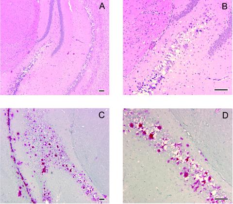
FIG. 1.
Neuropathology of Tg(CerPrP)1536+/− mice inoculated with CWD prions. Brains of sick animals from each study group were dissected rapidly after sacrifice and immersion fixed in 10% buffered paraformaldehyde. Tissue was embedded in paraffin, and sections were prepared and stained with hematoxylin and eosin for evaluation of spongiform degeneration. (A and B) Hematoxylin-and-eosin staining of sections through the hippocampus of Tg(CerPrP)1536+/− mice inoculated with brain tissue from CWD-affected mule deer D10 showing spongiform degeneration. Panel B is a higher magnification of an area in panel A. Note shrunken, scalloped neuronal nuclei adjacent to foci of spongiform change. (C and D) Immunohistochemistry of an adjacent section from the same inoculated Tg(CerPrP)1536+/− mouse showing amyloid plaque deposits. Panel D is a magnification of the area indicated in panel C. Note large immunoreactive plaques bordered by vacuoles. Slides were deparaffinized and hydrated followed by immersion in 88% formic acid solution, treatment with 25-mg/ml proteinase-K solution at 26°C for 10 min, followed by autoclaving for 20 min at 121°C in Tris-buffered solution. Tissue preparations were stained with anti-PrP polyclonal antibody R505 (8), followed by anti-rabbit immunoglobulin G-biotinylated secondary antibody streptavidin conjugated to alkaline phosphatase, and then developed with Fast Red A, naphthol, and Fast Red B chromogen. Hematoxylin was used as counterstain. Bar = 100 μm in all cases.

