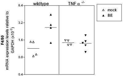
FIG. 8.
Expression of F4/80 mRNA following BE virus infection. Brain tissue was removed from BE virus-infected wild-type and TNF-α−/− mice at 21 days postinfection and analyzed for F4/80 mRNA as described for Fig. 7. Each symbol represents a single animal. Statistical analysis was performed by one-way analysis of variance with the Newman-Keuls posttest. The P value between mock-infected and BE virus-infected wild-type mice was <0.01, while the P value between mock-infected and BE virus-infected TNF-α−/− mice was >0.05.

