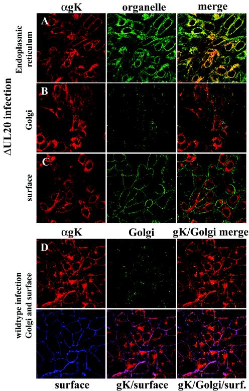
FIG. 3.
Digital images of confocal micrographs showing ΔUL20/gKDIV5 (A, B, and C) or gKDIV5/UL20amFLAG (D) virus-infected Vero cells. Infected cells were fixed at 12 hpi and stained with anti-V5 antibodies for gK (red) or specific organelle markers (green and blue) that specifically recognized ER (A), Golgi (green) (B and D), or plasma membranes (green in panel C, blue in panel D). Magnification, ×63; zoom, ×2.

