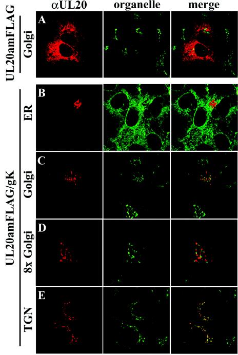
FIG. 6.
Digital images of confocal micrographs depicting UL20p localization in either UL20p-transfected (A) or UL20p- and gK-cotransfected (B to E) Vero cells. Transfected cells were fixed at 24 h posttransfection and stained with anti-FLAG antibodies for UL20p (red) or organelle markers (green) that specifically recognized ER (B), Golgi (A, C, and D), or TGN (E). Magnification, ×63; zoom, ×4 (A, B, C, and E) or ×8 (D).

