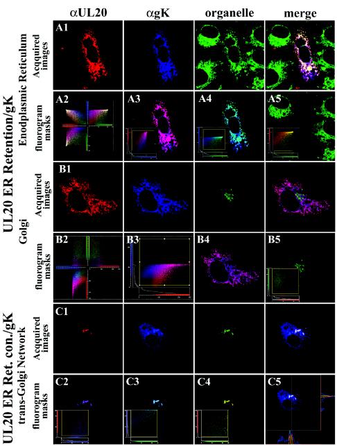
FIG.8.
Requirement for UL20p transport from ER to TGN for specific colocalization of gK, UL20p, and TGN markers. Vero cells coexpressing gK (green) and either the ER retention motif protein UL20p(KKSL) (A and B) or the ER retention control motif protein UL20p(KKSLAL) (C) were fixed at 24 h posttransfection, and the subcellular distributions of gK (blue) and UL20p (red) were determined relative to subcellular organelle markers (green) that specifically recognized ER (A), Golgi (B), or TGN (C). Three-dimensional fluorograms that depicted pixel intensity, distribution, and colocalization are shown for UL20p(KKSL) image subsets (panel 2 in A and B). Two-dimensional fluorograms and local image correlation masks between two fluorophores in each subset are shown for gK and UL20p(KKSL) (A, panel 3, and B, panels 3 and 4); gK and ER (A, panel 4); UL20p(KKSL) and Golgi (B, panel 5); UL20p(KKSLAL) and gK (C, panel 2); UL20p(KKSLAL) and TGN (C, panel 4); gK and TGN (C, panel 3). In addition, the region where no significant colocalization of gK, UL20p(KKSL), and ER occurred was masked (A, panel 5 inset), and as expected the relative image of this masked generated depicts no ER, gK, or UL20p(KKSL) proteins present. A sectional fluorohistogram for UL20p(KKSLAL), gK, and TGN colocalization is presented (C, panel 5) to show that all three markers specifically colocalized within most subcellular regions, except a few regions where only gK and UL20p(KKSLAL) colocalized outside of the TGN, most likely within Golgi membranes. Magnification, ×63; zoom, ×4.

