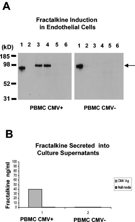
FIG. 1.
Fractalkine induction in endothelial cell cultures associated with CMV antigen-stimulated PBMC from CMV-seropositive donors. The data are representative of three separate experiments with different blood donors. (A) Detection of fractalkine induction in endothelial cell monolayers cocultured with CMV-seropositive PBMC by Western blot analysis with fractalkine-specific antibody. Lanes in left gel panel: 1, positive control for fractalkine, with ∼85-kDa form of fractalkine (note that the fractalkine form [R&D] lacks 57 carboxy-terminal amino acids and migrates at ∼85 kDa, whereas the endothelial-cell-associated form [also from R&D] is full length and migrates at ∼100 kDa); 2, negative control for fractalkine with endothelial cells in a resting state; 3 and 4, replicate samples of CMV antigen-treated PBMC from a CMV-seropositive donor; 5 and 6, replicate samples of media alone-treated PBMC from a CMV-seropositive donor. Lanes in right gel panel: 1, positive control for fractalkine, with ∼85-kDa form of fractalkine (commercially available from R&D); 2, negative control for fractalkine with endothelial cells in a resting state; 3 and 4, replicate samples of CMV antigen-treated PBMC from a CMV-seronegative donor; 5 and 6, replicate samples of medium-alone-treated PBMC from a CMV-seronegative donor. The arrow at the right of the gel indicates the Mr of fractalkine. (B) Detection of secreted fractalkine levels in coculture supernatants. ELISA results from coculture supernatants, comparing CMV-seropositive PBMC to CMV-seronegative PBMC.

