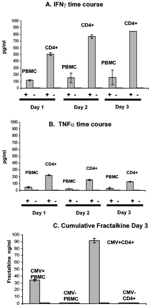
FIG. 6.
Time course in CMV antigen-stimulated CD4+ lymphocytes and PBMC cocultured with endothelial cells, showing cumulative levels of TNF-α (A) and IFN-γ (B) in culture supernatants over the course of 3 days. The results are shown with a CMV-seropositive donor, since the CMV-seronegative donor was completely negative for fractalkine induction. (C) Cumulative fractalkine levels by day 3 from same cocultures, showing results from a CMV-seropositive donor and a CMV-seronegative donor as a control. The data are representative of three separate experiments with different donors. The “+” or “−” symbol below graphs refer to samples with or without CMV antigen stimulation, respectively.

