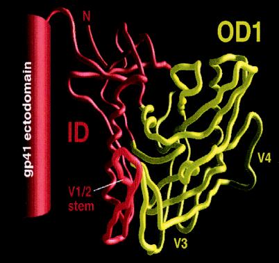
FIG. 1.
Schematic illustration of the OD1 and ID proteins. The Cα tracing of the gp120 core of HIV-1YU2 is derived from the structure of the CD4-gp120 core-17b Fab complex (35). The gp120 core structures comprising the OD1 protein are colored yellow; the hypothetical location of the V3 loop, which is missing from the gp120 core, is shown. The ID protein (red) includes the ID of the gp120 core and the gp120 V1/V2 loops (not shown), the C1 and C5 regions that are not in the gp120 core, and the gp41 ectodomain.

