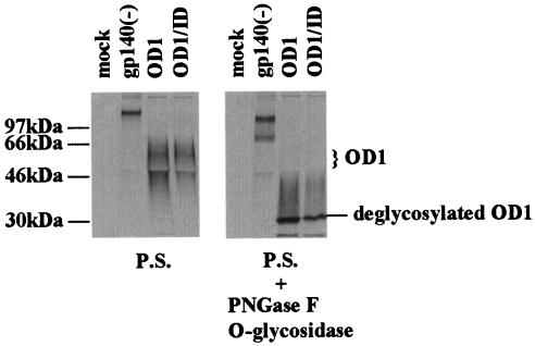
FIG. 2.
Expression of OD1 proteins in 293T cells. 293T cells transfected with a total of 10 μg of envelope glycoprotein expression plasmids were radiolabeled with [35S]cysteine-methionine, and the supernatants were incubated with a mixture of sera from HIV-1-infected persons (P.S.). The precipitates were analyzed on SDS-10% polyacrylamide gels. For OD1 and ID coexpression, 5 μg of each expressor plasmid DNA was transfected. Different species of the OD1 proteins are labeled on the right, and the molecular mass markers are indicated on the left. For deglycosylation, the protein-antibody-Protein A-agarose beads were boiled for 5 min in 1× sample buffer without β-mercaptoethanol. After incubation on ice for a few minutes, 500 U of PNGase F (New England Biolabs) and 0.5 mU of O-glycosidase (Boehringer Mannheim) were added to each sample, followed by incubation for 30 min in a 37°C water bath. The sample was boiled for 5 min before loading on a SDS-10% polyacrylamide gel.

