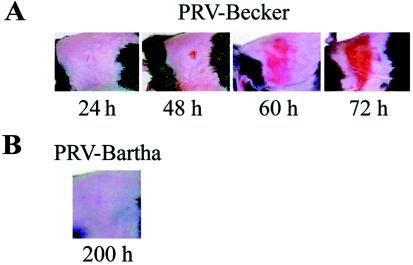
FIG. 1.
(A) Development and progression of the erythematous dermatome lesions observed in virulent PRV mouse flank infections. Mice were infected as described in Materials and Methods with PRV-Becker. Representative images of mice at 24, 48, 60, and 72 h are depicted. (B) Representative image of a PRV-Bartha-infected mouse at 200 h.

