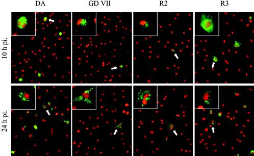
FIG. 2.
Detection of viral capsid antigens by immunofluorescence. Cell nuclei were counterstained with ethidium homodimer 1 (red). A representative field, observed at an original magnification of 25×, is shown for each viral strain. One representative infected cell, identified by a white arrow, is shown at higher magnification (×63) in the upper left quadrant to show the difference in intracellular distribution of the viral capsid proteins. Longer exposure times were needed to photograph the R2- and GDVII-infected samples; therefore, fluorescence intensities cannot be compared directly.

