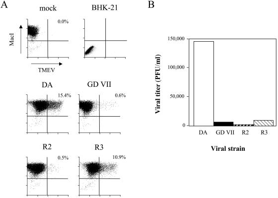
FIG. 4.
Analysis of TMEV-infected macrophage primary cultures by flow cytometry. Macrophages were seeded on six-well plates and infected at a multiplicity of infection of 5 PFU/cell. (A) Cells were harvested 24 h p.i. with Dispase II, fixed, permeabilized, and labeled with an anti-TMEV rabbit serum and a biotinylated rat monoclonal anti-MacI (CD11b) antibody. (B) The medium was harvested 24 h p.i. for viral titration.

