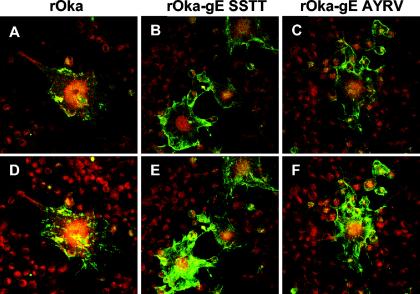
FIG. 2.

Localization of gE in melanoma cells 24 h after infection with rOka or rOka gE C-terminal mutants. Melanoma cells were infected with rOka (left panels), rOka-gE-SSTT (center panels), or rOka-gE-AYRV (right panels) for 24 h, and then the Golgi apparatus in live melanoma cells was pulse-labeled for 30 min with BODIPY Texas Red-ceramide complexed with BSA. After fixation, gE was detected with MAb 3B3 and FITC-conjugated anti-mouse IgG by confocal microscopy. Merged images from a single central plane (A, B, C) and merged projections spanning the entire depth of the monolayer (D, E, F) were obtained by using Adobe Photoshop software. Magnification, ×40.
