
FIG. 5.
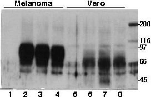
Analysis of gE maturation in melanoma and Vero cells infected with rOka and gE C-terminal mutants. Melanoma cells were mock infected (lane 1) or inoculated with rOka (lane 2), rOka-gE-SSTT (lane 3), or rOka-gE-AYRV (lane 4) and harvested after 72 h. Cell lysates were separated by SDS-10% PAGE and transferred to a nitrocellulose membrane; the mouse anti-gE MAb 3B3 was used as the probe, the secondary antibody was anti-mouse IgG conjugated with horseradish peroxidase, and the proteins were visualized by ECL. In parallel experiments, Vero cells were either mock infected (lane 5) or inoculated with rOka (lane 6), rOka-gE-SSTT (lane 7), or rOka-gE-AYRV (lane 8) and harvested after 72 h. Molecular markers that were radiolabeled with 14C are shown in the rightmost lane.