
Fig 1.
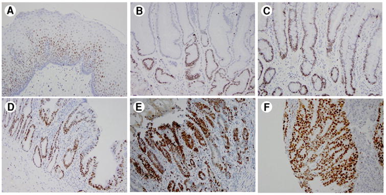
Immunohistochemical analysis of MCM4 in esophageal adenocarcinoma and precancerous lesions. A, Squamous mucosa. B, Columnar cell metaplasia. C, Barrett's esophagus. D, Low-grade dysplasia. E, High-grade dysplasia. F, Esophageal adenocarcinoma. In normal mucosa and non-dysplastic lesions, MCM4 nuclear staining is distributed in the basal layer of the epithelium and lower part of the glands. In dysplastic and cancerous lesions, the glands have full thickness staining for MCM4.