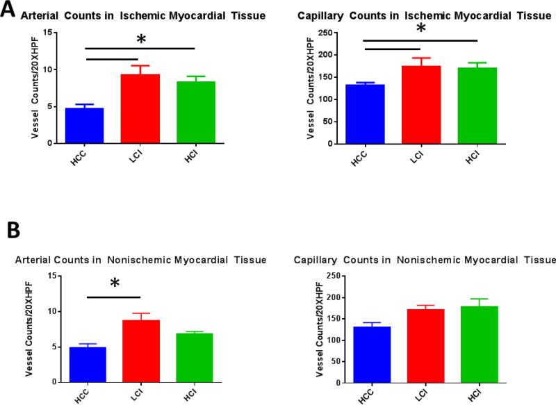
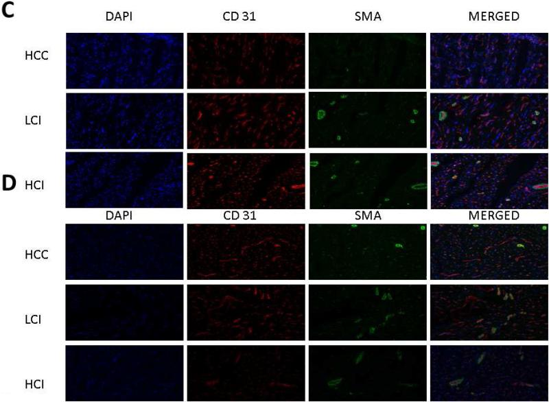
Figure 2. Calpain Inhibition Increases Vessel Density in Ischemic and Non Ischemic Myocardial Tissue.
A. Ischemic Myocardium: Arteriolar cell density staining for smooth muscle actin (SMA) with SMA specific antibody in tissue sections. Capillary cell density staining for endothelial marker CD31 with CD31 specific antibody in tissue sections. The bar diagrams show significant increases in LCI and HCI groups compared to control. B. Non-Ischemic Myocardium: Arteriolar cell density staining for smooth muscle actin (SMA) with SMA specific antibody in tissue sections. Capillary cell density staining for endothelial marker CD31 with CD31 specific antibody in tissue sections. The bar diagrams show an increase in arteriolar density in the LCI compared to control C. Ischemic Myocardium: Representative images in 20XHPF: CD31 is red. SMA is green. D: Non-Ischemic Myocardium: Representative images in 20XHPF: CD31 is red. SMA is green. HCC- High Cholesterol Control, LCI- Low Dose Calpain Inhibitor, HCI- High Dose Calpain Inhibitor; *= p<0.05 by Kruskal-Wallis and Dunn's Post Hoc Comparisons


