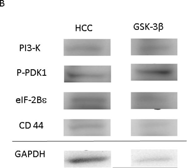
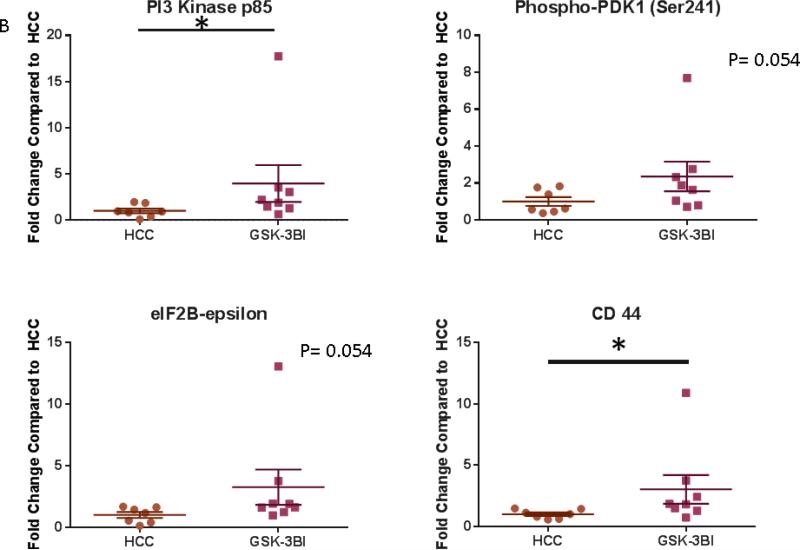
Figure 8. GSK-3β Increases Expression of Proteins involved Cell Growth and the WNT/ β Catenin Pathway.
A: Representative Images from Western Blot with protein specific antibodies as shown at the left. All images shown in for each protein are from the same Western blot membrane using different antibodies as indicated. GAPDH shows representative images for loading control. B: The bar diagrams show significant changes in pro survival signaling proteins. PI3Kp85- Phosphoinositide 3-Kinase, p-PDK-1 – Phospho-Phosphoinositide-dependent protein kinase 1 (ser 241) HCC- High Cholesterol Control, GSK-3βI- GSK-3β inhibited group *= p<0.05 by Mann-Whitney U-test


