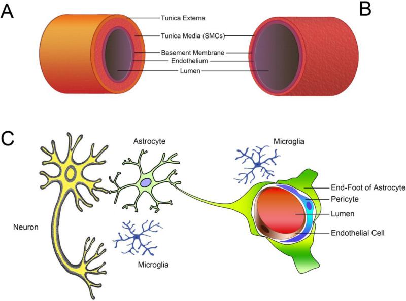
Fig. 1. Anatomical structures of the cerebral vasculature.
(A) Brain artery branch from the circle of Willis. The vessel wall contains three layers. The tunica externa is composed of connective tissue and elastic fibers. The tunica media is a thick layer of smooth muscle cells that control vessel tone. In the inner layer, the tunica intima consists of a monolayer of endothelial cells that rest on the basement membrane. (B) Brain arteries branch into a dense network of arterioles within the pia mater. These arterioles lack the tunica externa but contain the thinner media layer. As the major switch that controls the CBF, these arterioles are exposed to both blood in the lumen and the CSF rushing through the subarachnoid space. (C) The structure of the NVU is unique to the brain. Pericytes are contractile cells sitting outside the endothelial monolayer. The endothelial cells and neurons are connected by astrocyte endfeet. Microglial cells migrate around the NVU. Complex cell-cell interactions exist within the NVU and are integrated to control CBF and cellular function. SMCs: smooth muscle cells.

