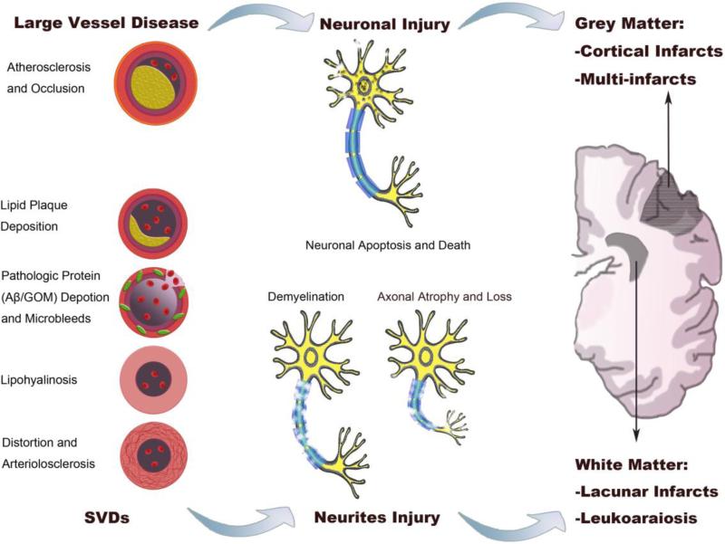
Fig. 2. Vascular and neural pathology underlying VCID.
The left panel represents diverse vascular pathologies in VCID. Large vessel diseases, including atherosclerosis and arterial occlusion, cause neuronal death (middle upper panel) or cerebral infarction. SVDs (left lower panel) typically cause neuritic injuries (middle lower panel), such as demyelination and axonal injuries. According to neuroimaging studies, VCID lesions can be observed in both gray and white matters (right panel). Among the lesions, cortical infarcts (typically multi-infarcts) are common in post-stroke VCID, while lacunar infarcts and leukoaraiosis are mainly located at periventricular regions. SVDs: small vessel diseases; Aβ: amyloid-β; GOM: granular osmiophilic material.

