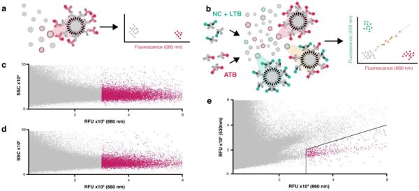
Figure 2. Serum IgG binding assay schematic and FACS-based high-throughput screening data.
(a) In single-color screens, library beads are incubated with serum-containing IgGs (gray IgGs), some of which bind specific beads. Probing with Alexa Fluor 647 anti-human IgG conjugate (red IgG, λem = 660 nm) labels serum IgG-bound beads for collection in FACS. (b) In two-color screens, serum pools are pre-labeled with anti-human mFab. The NCL pool is labeled with anti-human mFab488 (green Fab, λem = 530 nm), the ATB pool is labeled with anti-human mFab647 (red Fab, λem = 660 nm), and the pools are mixed. Probing the library with this mixed serum generates three populations of IgG-bound beads in multiplexed FACS analysis: NCL-specific IgG binding correlates with 530-nm fluorescence (upper left quadrant), ATB-specific IgG binding correlates with 660-nm fluorescence (lower right quadrant), and non-specific IgG and mFAB binding correlates with both channels (diagonal). FACS analysis of a single-color of pooled NCL serum screen (c) and pooled ATB serum screen (d) was gated for collection of hits > 30,000 RFU (λem = 660 nm, red). Side scatter (SSC) was used to gate single beads. Two-color FACS analysis of a mixed, pre-labelled NCL/ATB serum pool screen (e) wastwo-dimensionally gated (black lines) for hit collection (red).

