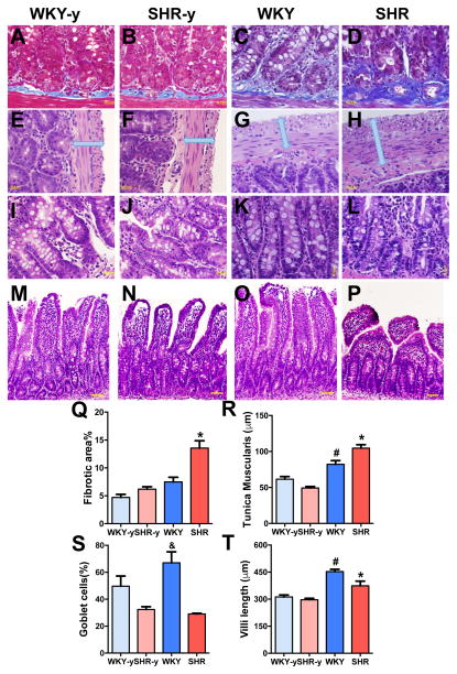
Figure 2. Pathological changes were observed in the small intestine of adult spontaneously hypertensive rat (SHR).
A–D, Small intestine was stained with Masson’s trichrome to quantify the fibrosis in young and adult WKY and SHR (Q, n=4/group). E–H, Cross sections of the small intestine of WKY and SHR were also stained with hematoxylin-eosin to measure the thickness of tunica muscularis layer (R). I–L, The number of goblet cells per 100 epithelial cells was decreased in SHR (S). M–P, Villi lengths were shorter in SHR (T). *p< 0.05 SHR vs all other groups; #p< 0.05 WKY vs all other groups; &p<0.05 WKY vs SHR-y and SHR.

