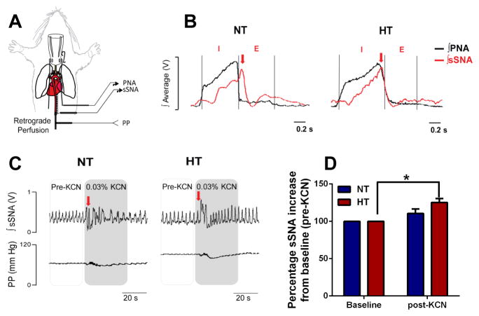
Figure 7. Elevated splanchnic sympathetic nerve activity in the SHR.
A, In situ decerebrated artificially-perfused rat preparation (schematics on the left) reveals respiratory uncoupling of the B, sSNA (red trace) in the hypertensive (HT) SHR compared to normotensive (NT) control rats. Phrenic nerve activity (PNA, black trace) inspiratory (I) and expiratory (E) phases were used to determine respiratory uncoupling. Note the typical shift in the peak of sSNA burst from the E phase in the NT (left panel, red arrow) to the I phase of PNA in the HT (right panel, red arrow). C–B, Intra-arterial bolus KCN injection produced a ~25% higher sSNA burst from baseline (pre-KCN) in the HT compared to NT rats (n=5/group). *p<0.05 baseline HT vs post-KCN HT.

