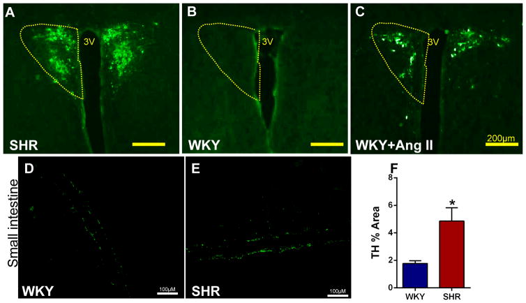
Figure 8. Higher PRV-GFP retrograde labeling from the small intestine to the PVN is associated with increased TH immunoreactivity in the small intestine of the SHR compared to WKY.
A–B, GFP staining reveals robust PRV retrograde labeling from small intestine to the PVN in the SHR compared to WKY. C, Retrograde labeling in WKY is enhanced following chronic Ang II-infusion. D–E, Representative images of tyrosine hydroxylase (TH) immunoreactivity in WKY and SHR small intestinal tissue. F, Quantification of TH staining revealed increased immunoreactivity in the small intestine of SHR (n=3/group). *p<0.05 SHR vs WKY.

