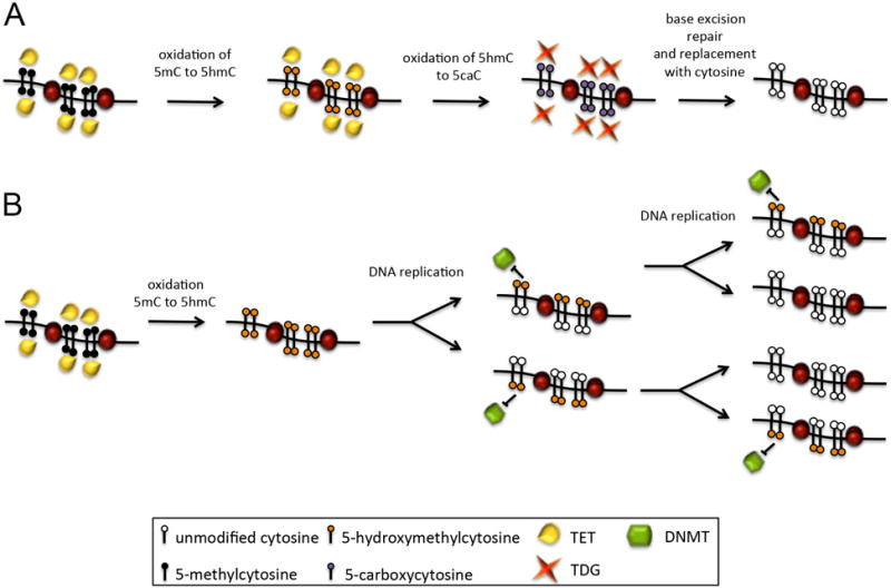
Figure 2. Models of demethylation mediated by the TET enzymes.

A) TET-assisted active demethylation. The TET enzymes sequentially oxidize methylated cytosines (5mC) into 5-hydroxymethylcytosine, then 5-formylcytosine (5fC) and finally 5-carboxycytosine (5caC). These altered cytosines can then be excised by the thymine DNA glycosylase enzyme (TDG) and replaced with an unmodified cytosine by part of the base-excision repair pathway. B) TET-assisted passive demethylation. The TET enzymes oxidize methylated cytosines into 5hmC, which are a poor substrate for DNMT activity after DNA replication. With each cycle of replication, the methylation status is further diluted.
