
FIG. 2.
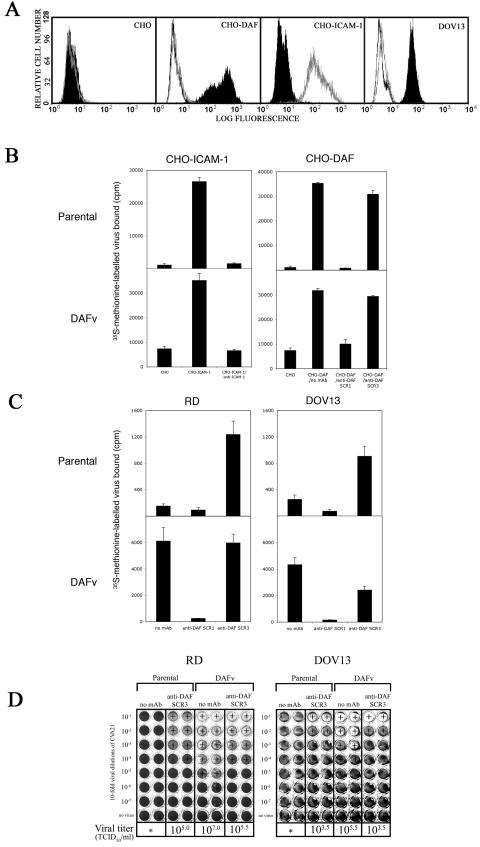
MAb blockade of CVA21-DAFv binding and lytic infection. (A) Flow cytometric analysis of surface levels of DAF and ICAM-1 on CHO, CHO-DAF, CHO-ICAM-1, and DOV13 cells. The open histogram represents binding of conjugate only, the dotted histogram represents binding of anti-ICAM-1 MAb, and the solid histogram represents binding of anti-DAF MAb. (B) Radiolabeled viral binding to surface-expressed ICAM-1 and DAF on transfected CHO cells measured by liquid scintillation counting. Results are expressed as the mean of triplicate samples plus standard deviation. (C) Radiolabeled viral binding to endogenously expressed DAF on RD and DOV13 cells measured by liquid scintillation counting. Results are expressed as the mean of triplicate samples plus standard deviation. (D) Effect of MAb cross-linking of DAF on CVA21 lytic infection of RD and DOV13 cells. Cell monolayers in 96-well plates were preincubated with anti-DAF SCR3 MAb prior to challenge with parental CVA21 and CVA21-DAFv (10-fold viral dilutions with 106 to 100 TCID50/well). Following incubation for 72 h at 37°C, the cell monolayers were fixed and stained with a crystal violet solution. Plus signs indicate a cytopathic effect detected by microscopic examination. Asterisks indicate a viral titer less than 10 TCID50/ml.