1.
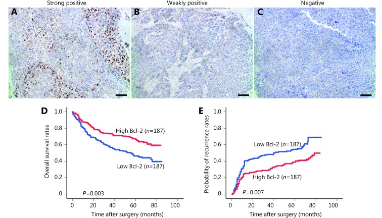
(A) Different expression status of Bcl-2 in tumor tissues detected by immunohistochemical staining and its correlation with the prognosis of HCC patients. (B) The graph was from a strong positive case in Bcl-2 expression. (C) The photograph shows a weekly positive expression level. (D) The graph was a totally negative one that showed little staining of Bcl-2. Positive cells were stained brown (100×); Bar = 100 μm. (E) High Bcl-2 density in tumor was associated with prolonged OS and TTR. (E) Different expression status of Bcl-2 in tumor tissues detected by immunohistochemical staining and its correlation with the prognosis of HCC patients.

