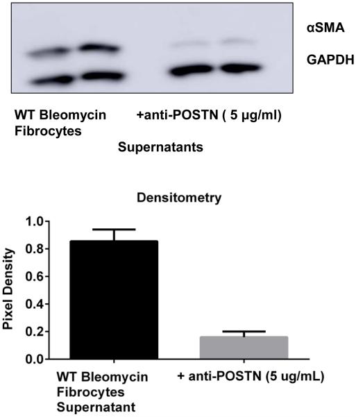
Figure 10. Neutralization of periostin secretion by fibrocytes post-bleomycin treatment limited their ability to promote myofibrolast differentiation.
WT mice were given 0.025U of bleomycin or PBS intratracheally on day 0. On day 14, post-bleomycin treatment lung mesenchymal cells were cultured for 14 days. After 14 days in culture cells were sorted and CD45 positive fibrocytes were incubated in SFM overnight in presence or absence of a mouse periostin neutralizing antibody (AF-2955, R&D systems). Cell-free supernatants were collected from both and added 1:1 with SFM onto WT untreated fibroblasts (CD45 negative) for 24h. Cells were lysed in RIPA buffer with protease inhibitor and we assessed the expression of αSMA and GAPDH by western blot. Data was quantitated using Image J software and pixel density is represented on bar graph.

